Temporal analysis of 3D data in nuclear medicine
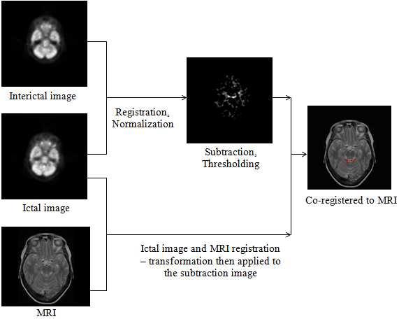
In nuclear medicine it is often needed to compare two tomographic scans, e.g. PET images of brain. It is useful to combine subtraction of the functional images with an anatomic image, e.g. MRI. One representative of such methods is SISCOM. It can be used to localize epileptogenic foci or to measure tumor development in time. The following image shows general scheme of such a method.
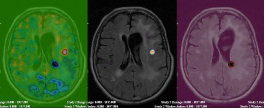
The next image shows difference image of a patient with tumor fused with MRI, its positive and its negative part.
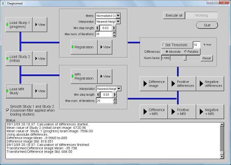
Based on a thorough comparison of available methods we developed a software to perform the subtraction task. The following figure shows its GUI. Other improvements will soon follow. We want to focus on subtraction of images with deformed tissue and of images taken with a new radiotracer FLT.
| Details: | |
| Duration: | 2008 - 2009 |
| Contact person: | Jiří Boldyš |
| Involved people: | Jan Kratochvíla (subject of his diploma thesis) |