Photorealistic Modeling of the Growth of Filamentous Specimens
| In this project, we introduce a method for modeling the growth of settled filamentous specimens in intervals between observations. The method is based on image warping and utilizes the properties of the morphological skeleton. |
 |
Objectives
Phytopathogenic fungi are able to cause severe diseases in a wide range of economically important crop plants. Detailed understanding of the pathogen development principles could contribute to the increasing efficiency of disease control.
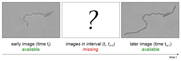
The microorganisms are examined mostly by means of light microscopy. As special conditions are required for their cultivation, their growth has to be documented repeatedly, in a defined sequence of observation sessions. Due to the complexity of the documentation process, the intervals are usually several hours long. In order to view the development of the specimen between two observation sessions, we have to reconstruct the missing images just from the images acquired at the beginning and at the end of the corresponding interval.
We restrict ourselves to settled specimens with filamentous growth patterns, such as some fungi and oomycetes. While their hyphae elongate over time, the growth in width, as well as movement of developed parts, is relatively negligible.


Light microscopy images of an early (left) and a later (right) stage of growth of Fusarium oxysporum f.sp. pisi, after preprocessing.
Methods
The method computes a mapping function that is utilized to warp the temporally closer of the available images to represent an arbitrary moment within the interval. The parameters of the geometric transformation are estimated by means of growth tracking based on the morphological skeleton. Due to the local character of the specimen's growth, elastic types of mapping functions are employed, e.g. radial basis functions called thin-plate splines.
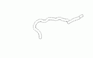
A sequence of control points computed from the available images, representing the shape of the specimen during the interval between observations.
Results
The result of image warping is a photorealistic artificial image of the specimen at the selected moment within the interval between observations.


An artificially generated image corresponding to 3/5 of the interval (left), and a reference image obtained for the purposes of comparison at the corresponding stage of growth (right).
Acknowledgment
The fungal strain UPOC-FUN-161 was provided by and its cultivation performed and documented at the Laboratory of Phytopathology, Department of Botany, Faculty of Science, Palacký University in Olomouc, Czech Republic.
| Details: | |
| Duration: | 2006-2008 |
| Funding: | GAUK 148207, MŠMT 1M0572, MSM 6198959215 |
| Contact person: | Jiří Sedlář |
| Involved people: | Jan Flusser |
| Involved extern: | Michaela Sedlářová, Palacký University in Olomouc |
Publications:
-
Photorealistic Modeling of the Growth of Filamentous Specimens, EURASIP Journal on Advances in Signal Processing vol.2008, 520972 (2008), p. 1-9
-
Tracking the Growth of Filamentous Fungi by Means of the Morphological Skeleton, Analysis of Biomedical Signals and Images. BIOSIGNAL 2006, p. 308-311, Eds: Jan J., Kozumplík J., Provazník I., Biosignal 2006, (Brno, CZ, 28.06.2006-30.06.2006)